Researchers from Harvard University and Google have created the most detailed map of human brain tissue ever produced.
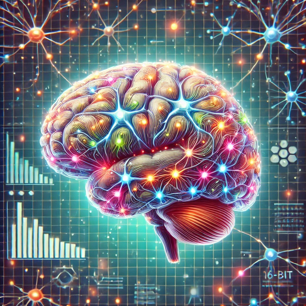
The team, led by Professor Jeff Lichtman, meticulously mapped a 1mm³ fragment of brain tissue taken from a patient undergoing epilepsy surgery. This seemingly small piece of brain contains approximately 57,000 cells and a staggering 150 million synapses, all reconstructed in 3D at nanoscale resolution.
This work took nearly 10 years of collaboration between Harvard’s Molecular and Cellular Biology department and Google’s connectomics research team. Combining electron microscopy and AI-powered algorithms, they were able to generate over 1.4 petabytes of data—a volume so vast it could be the foundation for new discoveries in neuroscience.
Interestingly, the research has already unveiled several unexpected findings. For example, they discovered neuron connections far stronger than typically observed in previous studies, with some neurons being connected by dozens of synapses.
Researchers believe these “super-strong” connections might play a role in speeding up information transfer within the brain. Moreover, odd structures such as axonal “whorls” have been found, although their role in brain function remains unclear.
Harvard and Google are not stopping here. Their future plans involve expanding this mapping project to include entire sections of the brain, with a specific focus on the hippocampal region, which is critical for memory and neurological diseases.

Leave a comment