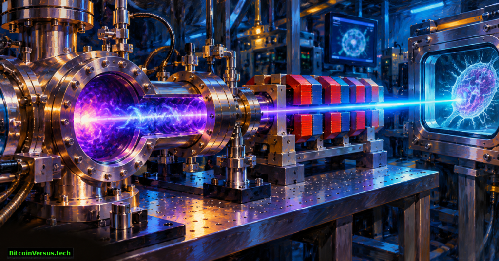
A newly published study proposes a compact free-electron laser capable of producing soft X-rays in a wavelength range useful for imaging biological cells, but the powerful output exists in computer simulations, not a completed experimental machine. Published November 5, 2026 in Nature Communications, the research models coherent X-ray pulses across the 2.3–4.4 nanometer “water window.” A team spanning the University of Strathclyde, Heinrich Heine University Düsseldorf and ELI Beamlines developed the approach.
The finding extends BitcoinVersus.Tech’s earlier account of a 420-nanometer laser-plasma experiment, which measured more than 1,000-fold optical gain and over 90% lasing-shot reliability. A separate follow-up documented more than eight hours of unattended operation. Both Berkeley Lab results involved actual light production. The new European proposal models different hardware and a much shorter wavelength.
Plasma physicist Dino Jaroszynski discusses laser-plasma accelerators and X-ray radiation sources in an earlier scientific lecture. Archival explanation, not a demonstration of the November 2026 simulations.
Why researchers want the water window
The water window covers approximately 2.3–4.4 nm. Water transmits soft X-rays in the range more readily than carbon-rich structures absorb them, creating useful contrast for observing cells and organic materials. Ordinary visible violet light at 420 nm cannot provide the same imaging mechanism. For scale, the proposed X-ray wavelengths are roughly 95–183 times shorter than 420 nm, but the comparison describes different accelerator designs rather than measured improvement to one machine.
Producing coherent radiation demands exceptionally bright, tightly controlled electron bunches. The proposed system first drives a plasma wake with an energetic electron beam. A precisely timed laser pulse then releases additional electrons inside the wake, acting as a plasma photocathode. Those newly released “witness” electrons accelerate through a metre-scale plasma stage.

The central challenge is electron-beam quality
The model adjusts witness-bunch charge to balance electric fields generated by the bunch against those of the plasma wake. Known as beam loading, the effect can reduce electron energy spread, which helps electrons remain synchronized as they pass through an undulator: alternating magnetic poles that force the beam into a gently oscillating path. Coherent light grows when emitted radiation reinforces subsequent emission.
The simulations project femtosecond-scale, multi-gigawatt peak X-ray pulses in a compact undulator. The authors also test sensitivity to changes in wakefield strength and photocathode-laser timing. Those are modeled tolerances, not measured operating stability, electricity consumption or commercial efficiency.
Carl B. Schroeder’s Berkeley Lab laser-plasma acceleration research talk describes particle acceleration and free-electron-laser applications. Historical technical context, not a validation of the Strathclyde simulation.
A promising design, not a working medical instrument
Heinrich Heine University says experimental testing is a future step. Realizing the concept would require a dependable high-brightness electron source, precise extraction and focusing, stable pulse timing, effective undulator integration and repeatable X-ray measurements. No prototype cost, wall-plug efficiency, imaging throughput or in-service lifetime was established by the published modeling.
A compact research source could eventually broaden access to radiation presently associated with large specialized facilities. Yet atomic-force microscopy, experimental nanolasers, and integrated photonic chips serve different kinds of measurements and must not be presented as interchangeable.
The verified advance is a new, peer-reviewed route for producing water-window X-rays in simulation. Experimental evidence remains the crucial milestone before claims about smaller facilities, broader access or industrial readiness can be tested.
Primary sources and media
- Berman et al., Nature Communications, November 5, 2026: original computational study
- Heinrich Heine University Düsseldorf, November 5, research announcement and original laboratory photography
- University of Strathclyde accepted research record, methods and publication dates
- Barber et al., July 2025: earlier experimental 420 nm optical-gain result
- Kohrell et al., April 2026: experimental extended operation, different system
- Jaroszynski technical talk and Schroeder Berkeley Lab talk
Editorial verification: The 2.3–4.4 nm range and multi-gigawatt pulse predictions describe numerical simulations. The published record does not document generated water-window X-rays. The featured oil painting is conceptual, the university photograph depicts a research laser facility, and historical lecture videos do not show the reported 2026 calculation.
Editor’s Note: We volunteer daily to ensure the credibility of the information on this platform is Verifiably True. If you would like to support to help further secure the integrity of our research initiatives, please scan the QR code below

For mobile scanning, open the article on another display and scan the QR code with a Bitcoin wallet. The voluntary donation amount is 5,000 sats. Verify the receiving address 3C9o19EH5HSiwEPyCTmEKzxhNCbo2X6TTb before sending.
BitcoinVersus.Tech publishes technology and financial journalism for informational purposes and does not provide financial, investment, or legal advice.


Leave a Reply